

Tissue Engineering ,The A-Z Comprehensive Guide
Luke McLaughlin, Biotech Digital Marketer, Business Developer and Life Science Content Creator

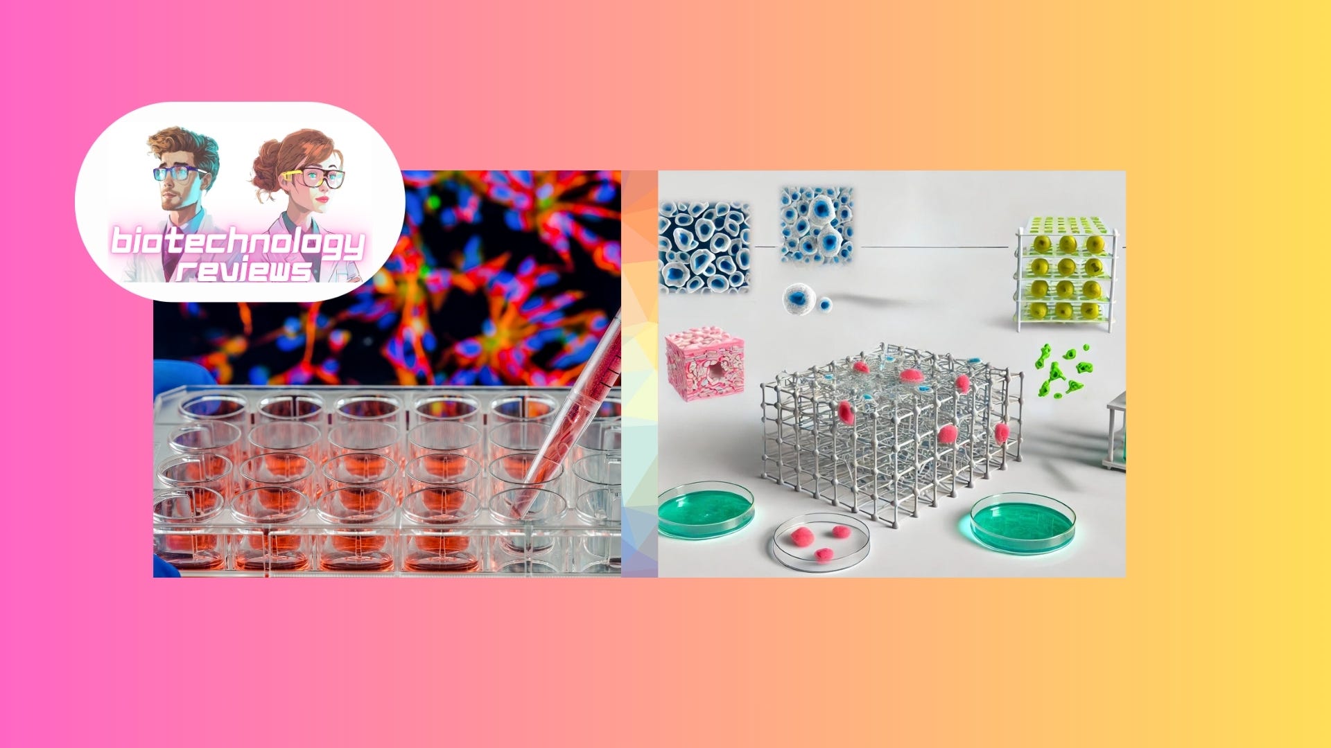
Tissue engineering is an advanced interdisciplinary field that merges principles from biology, engineering, and materials science with the aim of developing biological substitutes to restore, maintain, or enhance tissue function. The potential applications of tissue engineering are vast, including regenerative medicine, drug testing, disease modeling, and personalized medicine. By leveraging the body's intrinsic ability to heal and regenerate, tissue engineering seeks to create viable, functional tissues that can replace damaged or diseased organs.
This guide delves into the fundamental concepts, methodologies, protocols, and techniques employed in tissue engineering. Key components such as cells, scaffolds, and bioreactors are explored in depth. Cells, the building blocks of tissues, can be sourced from the patient (autologous), another human donor (allogeneic), or even different species (xenogeneic). Scaffolds are three-dimensional structures that support cell attachment, proliferation, and differentiation, providing the necessary mechanical and biochemical environment for tissue formation. Bioreactors, on the other hand, provide a controlled environment for tissue cultivation, simulating physiological conditions by regulating parameters such as temperature, pH, oxygen concentration, and mechanical forces.
Furthermore, this guide covers the detailed processes of cell extraction, isolation, and culture, ensuring the availability of high-quality, functional cells for tissue development. Advanced scaffold fabrication techniques such as electrospinning, 3D bioprinting, and decellularization are discussed, alongside the key properties and functionalization of scaffolds to enhance their performance in tissue engineering applications.
The role of bioreactors in enhancing tissue development and maturation is also examined, detailing various types and designs, including perfusion bioreactors, mechanical stimulation bioreactors, and rotating wall vessel bioreactors. Key parameters such as oxygen concentration, nutrient supply, and waste removal are critical for optimizing tissue culture conditions.
In addition to these core techniques, the guide explores advanced methods and emerging trends such as gene editing with CRISPR-Cas9, TALENs, and Zinc Finger Nucleases. These tools allow precise modification of cellular DNA to enhance cell properties, correct genetic defects, or introduce new functions, thereby expanding the possibilities for tissue engineering.
Finally, the guide introduces organ-on-a-chip technology, a cutting-edge approach that mimics the physiological functions of human organs on a microscale platform. This technology provides a more accurate representation of human organ function compared to traditional in vitro models, improving the predictive power of drug testing and disease modeling.
Through understanding and optimizing each step of these processes, researchers can enhance the efficacy and reliability of tissue engineering applications, pushing the boundaries of regenerative medicine and bringing us closer to the goal of creating functional, transplantable tissues and organs.
Contents
Introduction to Tissue Engineering
Fundamental Concepts
o Cells
Autologous Cells
Allogeneic Cells
Xenogeneic Cells
o Scaffolds
Natural Materials
Synthetic Materials
o Bioreactors
Cell Sourcing and Culture
o Cell Extraction and Isolation
Enzymatic Digestion
Mechanical Disruption
o Cell Isolation Techniques
Density Gradient Centrifugation
Magnetic-Activated Cell Sorting (MACS)
Fluorescence-Activated Cell Sorting (FACS)
o Cell Culture Techniques
Primary Cell Culture
Stem Cell Culture
Scaffold Fabrication
o Electrospinning
o 3D Bioprinting
o Decellularization
Bioreactors
o Types and Designs
Perfusion Bioreactors
Mechanical Stimulation Bioreactors
Rotating Wall Vessel Bioreactors
o Key Parameters
Advanced Techniques and Emerging Trends
o Gene Editing
CRISPR-Cas9
TALENs
Zinc Finger Nucleases (ZFNs)
o Applications in Tissue Engineering
Enhancing Cell Properties
Disease Modeling and Drug Testing
Regenerative Medicine
Organ-on-a-Chip Technology
o Design Principles
o Fabrication Techniques
Microfabrication
3D Printing
o Microfluidic Systems
o Organ-on-a-Chip Models
Lung-on-a-Chip
Heart-on-a-Chip
Liver-on-a-Chip
o Integration of Sensors
Types of Sensors
Integration Techniques
o Applications and Advantages
Drug Development
Disease Modeling
o Challenges and Future Directions
Fundamental Concepts
Cells: The essential units of life, cells form the basis of tissue engineering. Depending on the application, cells can be derived from various sources:
Autologous Cells: Derived from the same individual. These cells reduce the risk of immune rejection but may require invasive procedures for extraction and time for expansion in vitro.
Allogeneic Cells: Sourced from a donor of the same species. They are more readily available but carry a higher risk of immune rejection and transmission of diseases.
Xenogeneic Cells: Obtained from a different species, typically used for research due to ethical and immunological concerns in clinical applications.
Scaffolds: These are three-dimensional structures that support cell attachment, proliferation, and differentiation. Scaffolds provide the necessary mechanical and biochemical environment for tissue formation. They can be composed of:
Natural materials: Such as collagen, fibrin, or hyaluronic acid.
Synthetic materials: Including polylactic acid (PLA), polyglycolic acid (PGA), and polycaprolactone (PCL).
Bioreactors: Devices that provide a controlled environment for the cultivation of tissues. They simulate physiological conditions by regulating parameters such as temperature, pH, oxygen concentration, and mechanical forces.
Core Components
Tissue engineering's core components are cells, scaffolds, and bioreactors. Each of these elements is crucial for creating viable tissue constructs. Let's explore these in greater technical detail.
Cell Sources:
Primary Cells: Directly isolated from tissues, retaining the original tissue's properties but having limited proliferation capacity and potential for dedifferentiation.
Stem Cells:
Embryonic Stem Cells (ESCs): Pluripotent cells derived from the inner cell mass of blastocysts, capable of differentiating into any cell type. They have ethical and regulatory challenges.
Adult Stem Cells: Multipotent cells, such as mesenchymal stem cells (MSCs) from bone marrow or adipose tissue, which can differentiate into a limited range of cell types.
Induced Pluripotent Stem Cells (iPSCs): Somatic cells reprogrammed to a pluripotent state by introducing specific transcription factors (e.g., Oct4, Sox2, Klf4, and c-Myc). iPSCs offer a patient-specific and ethically favorable alternative to ESCs.
Cell Culture Techniques:
Isolation: Enzymatic digestion (e.g., trypsinization) and mechanical disruption are common methods for isolating cells from tissues.
Expansion: Culturing cells in vitro to increase their numbers involves using nutrient-rich media, growth factors, and maintaining optimal conditions (temperature, CO2 concentration).
Differentiation: Directed differentiation of stem cells into specific cell types using growth factors, cytokines, and physical cues. For instance, transforming growth factor-beta (TGF-β) for chondrogenic differentiation of MSCs.
Understanding the detailed processes of cell extraction, isolation, and culture is crucial for effective tissue engineering. These processes ensure that viable, functional cells are available for developing engineered tissues. Let's dive deeper into these techniques.
Cell Extraction and Isolation
Cell Extraction from Tissues
Enzymatic Digestion:
Proteolytic Enzymes: Commonly used enzymes include trypsin, collagenase, and dispase. These enzymes break down the extracellular matrix (ECM) to release cells.
Trypsin: A serine protease that cleaves peptide bonds at the carboxyl side of lysine and arginine residues. It's typically used in a 0.25% solution with EDTA to chelate calcium and magnesium, which enhances cell detachment.
Collagenase: Degrades collagen fibers in the ECM, facilitating the release of cells embedded within the matrix. Concentrations range from 0.1% to 0.5%, depending on tissue density.
Dispase: A neutral protease that cleaves fibronectin and collagen IV, preserving cell surface proteins, making it useful for gentle cell dissociation.
Mechanical Disruption:
Mincing: Physically cutting the tissue into small pieces to increase the surface area exposed to enzymatic digestion.
Homogenization: Using devices like blenders or tissue grinders to mechanically break down tissue, often followed by filtration to separate cells from debris.
Protocols:
Preparation: Sterilize instruments and prepare enzyme solutions.
Dissection: Mince the tissue into small fragments.
Enzymatic Treatment: Incubate tissue fragments in enzyme solution at 37°C with gentle agitation.
Filtration: Pass the digested tissue through a cell strainer (70 µm or 100 µm) to separate cells from undigested tissue.
Centrifugation: Collect cells by centrifuging at 300-500 g for 5-10 minutes, then resuspend in culture medium.
Cell Isolation Techniques
Density Gradient Centrifugation:
Ficoll-Paque: A commonly used density gradient medium for isolating mononuclear cells from blood. Blood is layered over Ficoll-Paque and centrifuged, resulting in distinct layers, with mononuclear cells forming a band at the plasma-Ficoll interface.
Percoll: Another medium used for gradient centrifugation, particularly for isolating cells based on size and density.
Magnetic-Activated Cell Sorting (MACS):
Uses magnetic beads conjugated to antibodies specific to cell surface markers. Cells labeled with these beads are passed through a magnetic column, where labeled cells are retained and unlabeled cells are washed away.
Positive Selection: Desired cells are labeled and retained.
Negative Selection: Unwanted cells are labeled and removed, leaving the desired cells in the flow-through.
Fluorescence-Activated Cell Sorting (FACS):
Cells are labeled with fluorescent antibodies specific to surface markers. Flow cytometry is used to sort cells based on fluorescence intensity.
Allows precise isolation of specific cell populations based on multiple markers.
Cell Culture Techniques
Primary Cell Culture
Medium Composition:
Basal Medium: Common formulations include DMEM (Dulbecco's Modified Eagle Medium), RPMI-1640, and MEM (Minimum Essential Medium).
Serum: Fetal bovine serum (FBS) is often added to provide growth factors, hormones, and attachment factors.
Antibiotics: Penicillin-streptomycin is added to prevent bacterial contamination.
Buffer Systems: HEPES or bicarbonate buffers maintain pH stability.
Culture Conditions:
Temperature: Typically 37°C for mammalian cells.
CO2 Concentration: 5% CO2 to maintain pH in bicarbonate-buffered media.
Humidity: 95% humidity to prevent medium evaporation.
Passaging:
Trypsinization: Cells are detached from the culture surface using trypsin-EDTA solution, then neutralized with serum-containing medium.
Splitting Ratios: Cells are reseeded at appropriate densities (e.g., 1:3 or 1:5) to maintain exponential growth.
Stem Cell Culture
Feeder Layers:
Mouse Embryonic Fibroblasts (MEFs): Used as a feeder layer for embryonic stem cells (ESCs) to provide essential growth factors.
Feeder-Free Systems: Utilize defined media supplemented with growth factors like bFGF (basic fibroblast growth factor) to maintain pluripotency.
Growth Factors and Supplements:
ESC Culture: Requires factors like LIF (leukemia inhibitory factor) for mouse ESCs or bFGF for human ESCs to maintain pluripotency.
MSC Culture: Often supplemented with factors like TGF-β, BMPs (bone morphogenetic proteins), or ascorbic acid to promote differentiation into specific lineages.
Differentiation Protocols:
Directed Differentiation: Involves the use of specific growth factors and signaling molecules to induce stem cells to differentiate into desired cell types.
Neural Differentiation: Using retinoic acid and noggin to induce neural progenitors.
Cardiomyocyte Differentiation: Activin A, BMP4, and bFGF to guide differentiation into cardiac cells.
Chondrogenic Differentiation: TGF-β3 and dexamethasone for cartilage formation.
Quality Control and Characterization
Viability and Proliferation Assays
Trypan Blue Exclusion: A dye exclusion test where viable cells exclude the dye, and dead cells take it up, allowing for viability assessment under a microscope.
MTT Assay: Measures cellular metabolic activity as an indicator of cell viability and proliferation.
Phenotypic Characterization
Immunocytochemistry: Staining cells with antibodies against specific markers to confirm cell identity and differentiation status.
Flow Cytometry: Analyzing cell surface markers to characterize cell populations and assess purity.
The detailed processes of cell extraction, isolation, and culture are fundamental to the success of tissue engineering. Mastery of these techniques ensures the availability of high-quality, functional cells for tissue development. By understanding and optimizing each step, researchers can enhance the efficacy and reliability of tissue engineering applications.
Scaffolds
Scaffolds are a critical component of tissue engineering, serving as the structural framework for cell attachment, proliferation, and differentiation. They mimic the extracellular matrix (ECM) and provide the necessary physical and biochemical environment for tissue development. This section delves into the materials, fabrication techniques, properties, and functionalization of scaffolds in technical detail.
Scaffold Materials
Natural Materials
Collagen:
Structure: Collagen is the most abundant protein in the ECM, providing tensile strength and structural support.
Types: Type I collagen is the most commonly used in scaffolding due to its high tensile strength.
Extraction: Collagen is typically extracted from animal tissues through acid or enzymatic digestion, followed by purification and lyophilization.
Crosslinking: To enhance mechanical stability, collagen scaffolds are often crosslinked using agents like glutaraldehyde, genipin, or through physical methods like UV irradiation.
Fibrin:
Formation: Fibrin is formed by the polymerization of fibrinogen in the presence of thrombin, resulting in a fibrous network.
Applications: Used in wound healing and tissue repair due to its excellent biocompatibility and hemostatic properties.
Degradation: Fibrin degrades naturally through fibrinolysis, mediated by plasmin.
Hyaluronic Acid:
Structure: A glycosaminoglycan composed of repeating disaccharide units of glucuronic acid and N-acetylglucosamine.
Properties: Highly hydrophilic, promoting cell migration and proliferation.
Modification: Hyaluronic acid can be chemically modified (e.g., methacrylation) to control its mechanical properties and degradation rate.
Synthetic Materials
Polylactic Acid (PLA):
Synthesis: PLA is synthesized through the ring-opening polymerization of lactide, a cyclic dimer of lactic acid.
Properties: Biodegradable, with a degradation rate that can be controlled by adjusting the polymer's molecular weight and crystallinity.
Applications: Used for bone and cartilage scaffolds due to its good mechanical strength and biocompatibility.
Polyglycolic Acid (PGA):
Synthesis: PGA is produced through the ring-opening polymerization of glycolide.
Properties: Rapidly biodegradable, making it suitable for applications where quick scaffold resorption is desired.
Applications: Commonly used in sutures and tissue engineering scaffolds for tissues that require rapid regeneration.
Polycaprolactone (PCL):
Synthesis: PCL is synthesized through the ring-opening polymerization of ε-caprolactone.
Properties: Slow degradation rate, good mechanical strength, and flexibility.
Applications: Ideal for long-term scaffold applications such as vascular grafts and nerve regeneration.
Scaffold Fabrication Techniques
Electrospinning
Principle: Electrospinning uses an electric field to draw a polymer solution or melt into ultra-fine fibers, creating a fibrous mat that mimics the ECM.
Process:
Solution Preparation: A polymer solution is prepared by dissolving the polymer in a suitable solvent.
Electrospinning Setup: The polymer solution is loaded into a syringe with a needle connected to a high-voltage power supply.
Fiber Formation: As the polymer solution is extruded from the needle tip, the electric field induces a charge on the solution, forming a Taylor cone. The charged jet elongates and thins due to electrostatic repulsion, forming fibers.
Collection: The fibers are collected on a grounded collector, forming a non-woven mat.
Parameters: Key parameters include the applied voltage (10-30 kV), flow rate (0.1-1 mL/h), distance between the needle and collector (10-20 cm), and polymer concentration.
3D Bioprinting
Principle: 3D bioprinting involves the layer-by-layer deposition of bioinks to create complex tissue structures with precise control over scaffold architecture and cell placement.
Techniques:
Extrusion-Based Bioprinting:
Process: Bioink is extruded through a nozzle to form continuous filaments.
Applications: Suitable for high-viscosity materials and creating large, mechanically robust structures.
Inkjet-Based Bioprinting:
Process: Droplets of bioink are deposited using thermal or piezoelectric actuation.
Applications: Ideal for low-viscosity bioinks and high-resolution printing.
Stereolithography (SLA):
Process: A photosensitive bioink is selectively polymerized layer-by-layer using a UV or visible light source.
Applications: Enables high-precision fabrication of intricate structures.
Bioinks: Common bioinks include alginate, gelatin methacrylate (GelMA), and decellularized ECM. These materials can be modified with cells, growth factors, and other biomolecules to enhance functionality.
Decellularization
Principle: Decellularization involves removing cellular components from tissues or organs while preserving the ECM structure, which can then be recellularized with patient-specific cells.
Methods:
Chemical Decellularization:
Detergents: SDS, Triton X-100, and CHAPS are commonly used to lyse cell membranes and solubilize cellular proteins.
Acid and Base Treatments: Acids like acetic acid and bases like sodium hydroxide can disrupt cellular components and remove nucleic acids.
Enzymatic Decellularization:
Proteases: Trypsin and dispase degrade cellular proteins.
Nucleases: DNase and RNase degrade nucleic acids, reducing the risk of immune response.
Physical Decellularization:
Freeze-Thaw Cycles: Repeated freezing and thawing cause ice crystal formation, which disrupts cell membranes.
High-Pressure Treatments: Applying high hydrostatic pressure can lyse cells while preserving the ECM.
Evaluation: The effectiveness of decellularization is assessed by evaluating residual DNA content, ECM composition, and structural integrity using histological staining, biochemical assays, and mechanical testing.
Scaffold Properties
Biocompatibility
Cell Adhesion: The scaffold surface must support cell attachment through integrin-mediated interactions with ECM proteins like fibronectin, laminin, and collagen.
Cytotoxicity: Materials should be non-toxic, as assessed by in vitro assays like the MTT or Alamar Blue assay.
Immunogenicity: Scaffolds should not elicit an adverse immune response. This is evaluated through in vivo implantation studies and immunohistochemical analysis.
Biodegradability
Degradation Rate: The scaffold should degrade at a rate that matches the rate of new tissue formation. This is controlled by the choice of material, polymer molecular weight, and crosslinking density.
Degradation Products: The by-products of scaffold degradation should be non-toxic and easily metabolized or excreted by the body.
Mechanical Properties
Strength and Stiffness: The scaffold must have sufficient mechanical strength to support the developing tissue and withstand physiological loads. Mechanical properties are tested using tensile, compressive, and dynamic mechanical analysis (DMA).
Elasticity: For tissues like skin and blood vessels, the scaffold should have appropriate elasticity to accommodate stretching and deformation.
Scaffold Functionalization
Surface Modification
Chemical Grafting:
Methods: Techniques like plasma treatment, chemical vapor deposition (CVD), and surface-initiated polymerization are used to graft functional groups onto the scaffold surface.
Applications: Enhancing cell adhesion, introducing bioactive molecules, and improving hydrophilicity.
Coating:
Methods: The scaffold surface can be coated with ECM proteins (e.g., collagen, fibronectin) or peptides (e.g., RGD) to enhance cell attachment and signaling.
Applications: Promoting specific cell functions, such as osteogenic or chondrogenic differentiation.
Incorporation of Growth Factors
Immobilization:
Methods: Growth factors like VEGF, BMPs, and FGF can be covalently bound to the scaffold surface or encapsulated within the scaffold matrix.
Release Kinetics: Controlled release of growth factors is achieved through the use of degradable linkers or encapsulating materials, ensuring sustained bioactivity over time.
Nanoparticle Delivery:
Methods: Growth factors are loaded into nanoparticles (e.g., PLGA, liposomes) that are incorporated into the scaffold. The nanoparticles release their cargo in response to environmental cues (e.g., pH, enzymatic activity).
Applications: Enhancing tissue regeneration, reducing inflammation, and promoting angiogenesis.
Scaffolds are integral to tissue engineering, providing the necessary support and environment for cell growth and tissue development. By carefully selecting materials, optimizing fabrication techniques, and functionalizing scaffolds with bioactive molecules, researchers can create sophisticated constructs that closely mimic the natural ECM. Continued advancements in scaffold technology hold the promise of developing functional, transplantable tissues and organs, revolutionizing regenerative medicine.
Bioreactors
Types of Bioreactors
Bioreactors play a crucial role in tissue engineering by providing a controlled environment that mimics physiological conditions, enhancing tissue development and maturation. They regulate parameters such as nutrient supply, waste removal, mechanical forces, and other environmental conditions essential for cell growth and differentiation. This section delves into the types, designs, functions, and operational principles of bioreactors in technical detail.
Types of Bioreactors
Perfusion Bioreactors
Principle: Perfusion bioreactors continuously perfuse culture medium through the scaffold, enhancing nutrient and oxygen delivery while removing metabolic waste products.
Design and Components:
Medium Reservoir: Holds the culture medium, which is often supplemented with nutrients, growth factors, and antibiotics.
Pump System: Circulates the medium through the scaffold. Common pump types include peristaltic pumps and syringe pumps.
Scaffold Chamber: Houses the scaffold where cells are seeded. It is designed to ensure uniform perfusion and minimal shear stress.
Sensors: Monitor key parameters such as pH, oxygen concentration, temperature, and pressure.
Tubing and Connectors: Sterile and biocompatible materials are used to connect the components and maintain a closed-loop system.
Operation:
Flow Rate: The perfusion flow rate is critical for maintaining optimal nutrient and oxygen levels. It is typically set based on the specific tissue type and scaffold properties.
Perfusion Modes: Continuous or intermittent perfusion modes can be used, depending on the requirements of the tissue being engineered.
Applications:
Bone Tissue Engineering: Enhances osteoblast proliferation and mineralization.
Liver Tissue Engineering: Supports hepatocyte function and detoxification.
Mechanical Stimulation Bioreactors
Principle: Apply mechanical forces to the developing tissue, promoting cellular alignment, proliferation, and differentiation. This mimics the physiological environment and mechanical loading experienced by tissues in vivo.
Design and Components:
Actuators: Devices that apply mechanical forces. These can be pneumatic, hydraulic, or motor-driven.
Force Transducers: Measure the magnitude of applied forces to ensure precise control.
Scaffold Holders: Secure the scaffold in place and transmit mechanical forces to the cells.
Types of Mechanical Stimulation:
Cyclic Compression: Applies compressive forces in a cyclic manner, typically used for cartilage and bone tissue engineering.
Tensile Stretching: Applies stretching forces to mimic the mechanical environment of tendons, ligaments, and muscles.
Shear Stress: Applies fluid-induced shear stress, important for vascular tissue engineering.
Operation:
Frequency and Amplitude: The frequency (e.g., 1 Hz) and amplitude (e.g., 10% strain) of mechanical loading are set based on the target tissue and experimental conditions.
Duration: Mechanical stimulation is applied for specific durations, often in cycles (e.g., 1 hour on, 1 hour off) to mimic natural loading patterns.
Applications:
Cartilage Tissue Engineering: Promotes chondrocyte proliferation and ECM production.
Tendon and Ligament Engineering: Enhances fibroblast alignment and collagen synthesis.
Rotating Wall Vessel Bioreactors
Principle: Create a low-shear, microgravity-like environment by rotating the culture vessel, promoting three-dimensional cell aggregation and tissue formation.
Design and Components:
Cylindrical Vessel: A horizontally rotating vessel that suspends cells in the culture medium.
Rotational Motor: Controls the speed of rotation, creating a dynamic environment.
Gas Exchange System: Ensures adequate oxygen supply and removal of carbon dioxide.
Operation:
Rotation Speed: The vessel's rotation speed (e.g., 10-20 rpm) is set to balance the forces, minimizing shear stress and sedimentation.
Culture Medium: The medium is continuously mixed, providing uniform nutrient distribution and waste removal.
Applications:
Stem Cell Aggregation: Promotes the formation of embryoid bodies from ESCs or spheroids from MSCs.
Tissue Constructs: Supports the development of complex tissues like cardiac or hepatic tissues.
Key Parameters in Bioreactor Operation
Oxygen Concentration
Importance: Critical for cell viability and function, especially for highly metabolic tissues like cardiac and hepatic tissues.
Monitoring and Control: Oxygen concentration is monitored using oxygen sensors. Control is achieved through gas exchange systems, typically using oxygen-permeable membranes or direct sparging.
Optimization: The optimal oxygen concentration depends on the specific cell type and tissue. Hypoxic conditions (1-5% O2) can be beneficial for some stem cell cultures, while others require normoxic conditions (21% O2).
Nutrient Supply
Medium Composition: The culture medium is supplemented with essential nutrients (e.g., glucose, amino acids, vitamins), growth factors, and hormones.
Flow Rate: The perfusion flow rate is optimized to ensure a continuous supply of nutrients while preventing accumulation of waste products.
Monitoring: Nutrient levels (e.g., glucose, glutamine) and metabolic by-products (e.g., lactate, ammonia) are monitored to adjust the medium composition and flow rate accordingly.
Waste Removal
Importance: Accumulation of metabolic waste products can be detrimental to cell growth and function.
Perfusion Systems: Continuous perfusion ensures efficient removal of waste products.
Medium Changes: In non-perfusion systems, periodic medium changes are performed to maintain optimal culture conditions.
Advanced Bioreactor Technologies
Microfluidic Bioreactors
Principle: Utilize microfluidic channels to provide precise control over the culture environment at a microscale level.
Design:
Microchannels: Small channels (10-500 µm) through which the culture medium flows.
Integrated Sensors: Real-time monitoring of pH, oxygen, and nutrient levels.
Micropumps: Control the flow rate of the medium.
Applications:
Organ-on-a-Chip: Mimics the microarchitecture and functions of human organs, providing a platform for studying tissue behavior and drug responses.
High-Throughput Screening: Enables the parallel testing of multiple conditions on a single chip.
Dynamic Culture Systems
Principle: Introduce dynamic conditions such as fluid flow, mechanical loading, or electrical stimulation to mimic the in vivo environment.
Design:
Dynamic Flow: Simulates blood flow or interstitial fluid flow.
Mechanical Loading: Applies cyclic mechanical forces.
Electrical Stimulation: Applies electrical pulses to promote the maturation of electrically active tissues like cardiac and neural tissues.
Applications:
Vascular Tissue Engineering: Promotes endothelial cell alignment and vessel formation.
Cardiac Tissue Engineering: Enhances cardiomyocyte maturation and synchronization.
Bioreactors are indispensable tools in tissue engineering, providing the controlled environments necessary for the development and maturation of engineered tissues. By understanding the intricacies of different bioreactor types, designs, and operational parameters, researchers can optimize tissue culture conditions, enhancing the quality and functionality of engineered tissues. Continued advancements in bioreactor technology will further drive the field of regenerative medicine, bringing us closer to the realization of functional, transplantable tissues and organs.
Tissue Engineering Methods and Techniques
In tissue engineering, the methods and techniques used for developing functional tissue constructs are diverse and complex. Below, we will explore the intricacies of cell sourcing and culture, scaffold fabrication, and bioreactor design in technical detail.
Cell Sourcing and Culture
Cell Isolation Techniques
Density Gradient Centrifugation:
Principle: This technique separates cells based on their density using a medium with a gradient of densities. The most common mediums used are Ficoll-Paque and Percoll.
Protocol:
Sample Preparation: Blood or tissue homogenate is diluted in a balanced salt solution.
Layering: The diluted sample is carefully layered over the density gradient medium in a centrifuge tube.
Centrifugation: The tube is centrifuged at a specific speed (e.g., 400-800 g) for 20-30 minutes without braking.
Harvesting: After centrifugation, distinct layers form, with the desired mononuclear cells at the interface between the plasma and the gradient medium. These cells are carefully aspirated and washed for further use.
Magnetic-Activated Cell Sorting (MACS):
Principle: Uses magnetic beads conjugated to antibodies specific to cell surface markers.
Protocol:
Labeling: Cells are incubated with magnetic beads coated with antibodies targeting specific cell surface antigens.
Separation: The cell suspension is passed through a magnetic column. Cells labeled with magnetic beads are retained in the column while unlabeled cells pass through.
Elution: The retained cells are then eluted by removing the column from the magnetic field and washing with buffer.
Fluorescence-Activated Cell Sorting (FACS):
Principle: Utilizes fluorescently labeled antibodies and flow cytometry to sort cells based on fluorescence intensity.
Protocol:
Labeling: Cells are stained with fluorescent antibodies specific to desired cell surface markers.
Flow Cytometry: The cell suspension is passed through a flow cytometer, where cells are individually analyzed for fluorescence.
Sorting: Based on fluorescence intensity, cells are sorted into different populations using electrostatic deflection.
Cell Culture Techniques
Primary Cell Culture:
Medium Composition:
Basal Medium: Compositions like DMEM, RPMI-1640, and MEM include essential nutrients, vitamins, and amino acids.
Serum: FBS is added to provide hormones, growth factors, and attachment proteins.
Supplements: Additional supplements such as glutamine, non-essential amino acids, and sodium pyruvate may be added based on specific cell needs.
Antibiotics: Penicillin-streptomycin is commonly used to prevent bacterial contamination.
Passaging Protocol:
Preparation: Pre-warm trypsin-EDTA solution and culture medium.
Trypsinization: Aspirate the old medium, wash cells with PBS, and add trypsin-EDTA solution to detach cells from the culture flask.
Neutralization: After cells detach (observed under a microscope), add serum-containing medium to neutralize trypsin.
Centrifugation: Collect the cell suspension and centrifuge at 300-500 g for 5 minutes.
Resuspension: Resuspend the cell pellet in fresh culture medium and seed into new culture flasks at the desired density.
Stem Cell Culture:
Feeder Layers:
MEFs: Typically inactivated by mitomycin C or gamma irradiation to prevent proliferation but continue to provide necessary growth factors.
Feeder-Free Systems: Use defined media such as mTeSR1 or TeSR-E8 supplemented with growth factors like bFGF to maintain pluripotency.
Differentiation Protocols:
Neural Differentiation: ESCs or iPSCs are treated with retinoic acid and noggin to induce differentiation into neural progenitors. Media is often supplemented with B27 and N2.
Cardiomyocyte Differentiation: Activin A and BMP4 are used to induce mesoderm formation, followed by bFGF and VEGF to promote cardiomyocyte differentiation.
Chondrogenic Differentiation: MSCs are cultured in high-density micromass or pellet cultures in medium supplemented with TGF-β3, dexamethasone, and ascorbic acid.
Scaffold Fabrication
Scaffold Materials
Natural Materials:
Collagen: Provides a natural ECM structure and promotes cell adhesion. It is often crosslinked to enhance mechanical strength and stability.
Fibrin: Forms a natural scaffold with excellent biocompatibility, used in wound healing and tissue repair.
Hyaluronic Acid: A glycosaminoglycan that provides a hydrated environment and promotes cell migration and proliferation.
Synthetic Materials:
Polylactic Acid (PLA): Biodegradable polymer with tunable mechanical properties, used for bone and cartilage scaffolds.
Polyglycolic Acid (PGA): Degrades rapidly, making it suitable for tissues that require quick scaffold replacement.
Polycaprolactone (PCL): A slowly degrading polymer with good mechanical strength, used for long-term scaffold applications.
Fabrication Techniques
Electrospinning:
Process: A polymer solution is fed through a needle and subjected to a high-voltage electric field, creating fine fibers collected on a grounded target.
Parameters: Voltage (10-30 kV), polymer concentration (5-20 wt%), flow rate (0.1-1 mL/h), and distance between the needle and collector (10-20 cm) are critical for controlling fiber diameter and morphology.
3D Bioprinting:
Bioinks: Hydrogels mixed with cells, such as alginate, gelatin methacrylate (GelMA), or decellularized ECM, are used as bioinks.
Printing Techniques:
Extrusion-Based: Continuous deposition of bioink through a nozzle, suitable for high-viscosity materials.
Inkjet-Based: Droplet-by-droplet deposition, suitable for low-viscosity materials.
Stereolithography (SLA): Uses light to polymerize a photosensitive bioink layer-by-layer.
Decellularization:
Chemical Methods: Use of detergents like SDS and Triton X-100 to lyse cells and solubilize cellular components while preserving the ECM.
Enzymatic Methods: Trypsin and nucleases (e.g., DNase, RNase) are used to degrade cellular components without damaging the ECM.
Physical Methods: Freeze-thaw cycles and high-pressure treatments to disrupt cell membranes and remove cellular debris.
Bioreactors
Types and Designs
Perfusion Bioreactors:
Principle: Medium is continuously perfused through the scaffold, enhancing nutrient and oxygen delivery while removing waste products.
Design: Typically includes a pump to circulate the medium, a scaffold chamber, and sensors for monitoring pH, oxygen, and temperature.
Mechanical Stimulation Bioreactors:
Principle: Apply mechanical forces to the developing tissue, promoting cellular alignment and tissue maturation.
Design: Includes actuators to apply cyclic compression, tension, or shear stress. Parameters like force magnitude, frequency, and duration are controlled.
Rotating Wall Vessel Bioreactors:
Principle: Creates a low-shear, microgravity-like environment by rotating the culture vessel, promoting three-dimensional cell aggregation and tissue formation.
Design: Consists of a cylindrical vessel that rotates around its horizontal axis, with cells suspended in the medium, minimizing shear stress.
Key Parameters
Oxygen Concentration: Critical for cell viability and function, especially for highly metabolic tissues. Controlled using oxygen sensors and gas exchange systems.
Nutrient Supply: Continuous supply of essential nutrients (glucose, amino acids, vitamins) and growth factors. Media composition tailored to the specific needs of the cells and tissue.
Waste Removal: Efficient removal of metabolic waste products like lactate and ammonia, typically achieved through perfusion systems or frequent media changes.
The detailed processes involved in cell sourcing and culture, scaffold fabrication, and bioreactor design are integral to the success of tissue engineering. Mastering these methods and techniques ensures the production of high-quality, functional tissue constructs. By continuously optimizing and innovating within these domains, researchers can push the boundaries of regenerative medicine and bring us closer to the goal of creating transplantable tissues and organs.
Advanced Tissue Engineering Techniques and Emerging Trends
Gene Editing
Techniques like CRISPR/Cas9 allow precise modification of cellular DNA, enabling the correction of genetic defects or the enhancement of cell properties for tissue engineering applications.
Gene Editing in Tissue Engineering
Gene editing is a powerful tool in tissue engineering, enabling precise modification of the genetic material within cells to enhance their properties, correct genetic defects, or introduce new functions. This section will cover the methodologies, mechanisms, applications, and challenges of gene editing in the context of tissue engineering in technical detail.
Gene Editing Technologies
CRISPR-Cas9
Principle: The CRISPR-Cas9 system is derived from a bacterial immune mechanism and allows for targeted DNA double-strand breaks (DSBs) at specific genomic locations.
Components:
Cas9 Nuclease: An enzyme that induces DSBs at the target site.
Guide RNA (gRNA): A synthetic RNA molecule that directs the Cas9 to the specific DNA sequence by base-pairing with the target genomic DNA.
Mechanism:
Design: gRNA is designed to match the target DNA sequence.
Binding: gRNA-Cas9 complex binds to the target DNA through the gRNA.
Cleavage: Cas9 induces a DSB at the target site.
Repair: The cell's natural DNA repair mechanisms, non-homologous end joining (NHEJ) or homology-directed repair (HDR), repair the break. NHEJ often results in insertions/deletions (indels), while HDR can introduce precise modifications if a repair template is provided.
Applications:
Gene Knockout: Disruption of specific genes to study their function or to remove detrimental genes.
Gene Correction: Introduction of correct sequences to repair genetic mutations.
Gene Insertion: Addition of new genes or regulatory elements to enhance cell properties or introduce new functions.
TALENs (Transcription Activator-Like Effector Nucleases)
Principle: TALENs are engineered nucleases that induce DSBs at specific DNA sites, similar to CRISPR-Cas9, but utilize a different DNA-binding mechanism.
Components:
DNA-Binding Domain: Composed of TALE repeats, each recognizing a single nucleotide.
FokI Nuclease: A dimeric nuclease that cleaves DNA when two TALEN monomers bind adjacent sites.
Mechanism:
Design: TALE repeats are engineered to recognize the target DNA sequence.
Binding: Two TALEN monomers bind to adjacent sites on the DNA.
Cleavage: FokI nuclease domains dimerize and induce a DSB.
Repair: The DSB is repaired by NHEJ or HDR, similar to CRISPR-Cas9.
Applications:
Gene Editing: Precise modification of specific genomic loci.
Functional Studies: Investigating the roles of specific genes in development and disease.
Zinc Finger Nucleases (ZFNs)
Principle: ZFNs are engineered nucleases that combine a DNA-binding domain of zinc finger proteins with the FokI nuclease to induce targeted DSBs.
Components:
Zinc Finger Proteins: Modular proteins that recognize specific 3-4 base pair DNA sequences.
FokI Nuclease: A dimeric nuclease that cleaves DNA when two ZFN monomers bind adjacent sites.
Mechanism:
Design: Zinc finger domains are engineered to bind specific DNA sequences.
Binding: Two ZFN monomers bind to adjacent sites on the DNA.
Cleavage: FokI nuclease domains dimerize and induce a DSB.
Repair: The DSB is repaired by NHEJ or HDR.
Applications:
Targeted Mutagenesis: Introduction of mutations or deletions at specific loci.
Gene Therapy: Correction of genetic defects in clinical applications.
Applications in Tissue Engineering
Enhancing Cell Properties
Stem Cell Engineering:
Pluripotency Maintenance: Introduction of pluripotency factors (e.g., OCT4, SOX2) to maintain or induce pluripotency in stem cells.
Differentiation: Knockout or overexpression of genes to direct stem cell differentiation into specific lineages (e.g., neural, cardiac).
Immune Evasion:
Gene Knockout: Deletion of major histocompatibility complex (MHC) genes to reduce immune recognition and rejection in allogeneic transplants.
Immune Modulation: Introduction of genes that encode for immunosuppressive factors (e.g., PD-L1) to evade immune detection.
Disease Modeling and Drug Testing
In Vitro Models:
Genetic Diseases: Introduction of disease-specific mutations into iPSCs to create patient-derived cellular models for studying disease mechanisms and testing therapies.
Drug Screening: Use of genetically modified cells to test the efficacy and toxicity of new drugs in a high-throughput manner.
Organ-on-a-Chip:
Tissue-Specific Mutations: Incorporation of gene-edited cells into organ-on-a-chip systems to model diseases in a tissue-specific context, allowing for more accurate drug response studies.
Regenerative Medicine
Gene Therapy:
In Situ Correction: Direct correction of genetic defects in target tissues using viral or non-viral delivery systems for gene editing tools.
Ex Vivo Editing: Gene editing of patient-derived cells followed by transplantation back into the patient to treat genetic disorders (e.g., beta-thalassemia, sickle cell anemia).
Tissue Repair and Regeneration:
Growth Factor Regulation: Introduction of genes encoding growth factors (e.g., VEGF, BMPs) to promote tissue regeneration and repair.
Anti-Apoptotic Genes: Introduction of genes that prevent cell death (e.g., BCL-2) to enhance the survival and function of transplanted cells.
Challenges and Considerations
Delivery Methods
Viral Vectors:
Adeno-Associated Virus (AAV): Efficient gene delivery with low immunogenicity, but limited cargo capacity.
Lentivirus: Capable of integrating into the host genome, providing long-term expression, but with a risk of insertional mutagenesis.
Non-Viral Methods:
Lipid Nanoparticles (LNPs): Deliver CRISPR components through cell membranes efficiently, with ongoing optimization for specificity and efficiency.
Electroporation: Uses electric pulses to transiently permeabilize cell membranes for gene delivery, effective for ex vivo editing but less so for in vivo applications.
Off-Target Effects
Specificity: Ensuring that gene editing tools target the intended genomic locus without affecting other regions. Off-target effects can lead to unintended mutations, potentially causing oncogenic transformations or other harmful effects.
Detection: Techniques like whole-genome sequencing (WGS) and GUIDE-seq are used to identify and quantify off-target effects, enabling the refinement of gene editing tools to improve specificity.
Ethical and Regulatory Issues
Ethical Considerations: Gene editing in human embryos and germline cells raises ethical concerns about heritable genetic changes and potential misuse for non-therapeutic enhancements.
Regulatory Approvals: Ensuring compliance with regulatory guidelines and obtaining approvals for clinical applications of gene editing technologies.
Gene editing is a transformative tool in tissue engineering, enabling precise manipulation of genetic material to enhance cell properties, model diseases, and develop advanced regenerative therapies. By understanding the mechanisms, applications, and challenges associated with gene editing technologies, researchers can optimize these tools for various tissue engineering applications, paving the way for innovative treatments and advancing the field of regenerative medicine.
Organ-on-a-Chip Technology
Organ-on-a-chip (OOC) technology is a cutting-edge approach in tissue engineering that seeks to mimic the physiological functions of human organs on a microscale platform. These systems provide a more accurate representation of human organ function compared to traditional in vitro models, thereby improving the predictive power of drug testing and disease modeling. This section delves into the technical aspects of OOC, including design principles, fabrication techniques, and applications.
Design Principles
Organ-on-a-chip devices aim to replicate the architecture and functionality of human organs. Key design considerations include:
Microarchitecture: Recreating the complex microenvironment of tissues, including cell organization, ECM composition, and vascularization.
Microfluidics: Using microfluidic channels to simulate blood flow and nutrient transport, providing dynamic conditions that mimic the in vivo environment.
Mechanical Stimulation: Incorporating mechanical cues such as shear stress, compression, and stretch to replicate the physical forces experienced by tissues.
Integration of Sensors: Embedding sensors to monitor physiological parameters in real-time, such as pH, oxygen levels, and metabolic activity.
Fabrication Techniques
Microfabrication
Soft Lithography:
Process: Soft lithography involves creating a master mold using photolithography, followed by casting a soft elastomer like polydimethylsiloxane (PDMS) to replicate the microfeatures.
Steps:
Photolithography: A silicon wafer is coated with a photoresist and exposed to UV light through a mask, creating a pattern on the wafer.
Development: The exposed photoresist is developed, revealing the patterned mold.
PDMS Casting: Liquid PDMS is poured onto the mold and cured, creating a flexible replica of the microfeatures.
Bonding: The PDMS layer is bonded to a glass or another PDMS layer to form sealed microchannels.
Applications: Soft lithography is widely used for creating microfluidic channels and compartments in OOC devices.
3D Printing
Additive Manufacturing:
Process: 3D printing involves layer-by-layer deposition of materials to build complex structures. Techniques such as stereolithography (SLA), fused deposition modeling (FDM), and inkjet printing are commonly used.
Materials: Biocompatible materials like hydrogels (e.g., GelMA), thermoplastics (e.g., PLA, PCL), and photopolymers are used to construct OOC devices.
Applications: 3D printing enables the fabrication of intricate designs, including vascular networks and multi-material constructs, enhancing the complexity and functionality of OOC systems.
Microfluidic Systems
Design and Components
Microchannels:
Function: Microchannels mimic the blood vessels and tissue interfaces, providing a controlled environment for fluid flow and nutrient transport.
Dimensions: Typical dimensions range from 10 to 500 micrometers in width and height, designed to match the scale of capillaries and small blood vessels.
Micropumps and Valves:
Function: Micropumps and valves control the flow rate and direction of fluids within the microchannels.
Types: Common types include peristaltic pumps, pneumatic pumps, and electrokinetic pumps.
Integration:
Connections: Microchannels are connected to external reservoirs and pumps via microfluidic tubing and connectors, ensuring a continuous supply of culture medium.
Interfacing: Devices often include interfaces for sensor integration, allowing real-time monitoring of physiological parameters.
Organ-on-a-Chip Models
Lung-on-a-Chip
Design:
Microarchitecture: Consists of a porous membrane separating two microchannels, with one side lined with lung epithelial cells and the other with endothelial cells.
Mechanical Actuation: Cyclic stretching mimics breathing movements, enhancing cell differentiation and function.
Applications:
Drug Testing: Evaluating the efficacy and toxicity of respiratory drugs.
Disease Modeling: Studying lung diseases like asthma, COPD, and pulmonary infections.
Heart-on-a-Chip
Design:
Microarchitecture: Incorporates microchannels lined with cardiac cells, often arranged in a specific orientation to mimic myocardial tissue structure.
Electrical Stimulation: Electrodes integrated into the device provide electrical pulses to induce cardiomyocyte contraction.
Applications:
Drug Screening: Testing cardiotoxicity and efficacy of cardiovascular drugs.
Disease Modeling: Investigating cardiac diseases like arrhythmias and cardiomyopathies.
Liver-on-a-Chip
Design:
Microarchitecture: Features multiple microchannels with hepatocytes and non-parenchymal cells, simulating the liver's multi-cellular environment.
Perfusion System: Continuous flow of culture medium mimics blood flow, supporting hepatic functions like metabolism and detoxification.
Applications:
Drug Metabolism: Studying the pharmacokinetics and pharmacodynamics of drugs.
Toxicology: Assessing liver toxicity of pharmaceuticals and environmental chemicals.
Integration of Sensors
Types of Sensors
Electrochemical Sensors:
pH Sensors: Monitor the acidity/alkalinity of the culture medium.
Oxygen Sensors: Measure dissolved oxygen levels to ensure adequate oxygen supply to cells.
Optical Sensors:
Fluorescence Sensors: Detect specific biomarkers or metabolites using fluorescent probes.
Optical Density Sensors: Measure cell growth and proliferation by detecting changes in optical density.
Integration Techniques
On-Chip Integration:
Embedded Sensors: Sensors are embedded directly into the OOC device during fabrication, allowing real-time monitoring of physiological parameters.
Microelectrodes: Electrodes are fabricated on-chip to measure electrical activity, particularly useful in heart-on-a-chip and neural-on-a-chip models.
Data Acquisition:
Interface Circuits: Interface circuits amplify and process sensor signals, converting them into digital data.
Software Integration: Software platforms collect and analyze sensor data, providing real-time feedback and control.
Applications and Advantages
Drug Development
High-Throughput Screening:
Parallel Testing: OOC platforms allow simultaneous testing of multiple drug candidates under physiologically relevant conditions.
Reduced Costs: Decreases the need for animal testing and reduces overall drug development costs.
Predictive Power:
Human Relevance: OOC models provide a more accurate representation of human organ responses compared to traditional cell culture and animal models.
Personalized Medicine: Patient-derived cells can be used to create personalized OOC models, enabling tailored drug treatments.
Disease Modeling
Complex Diseases:
Multi-Organ Chips: Combining multiple OOC models (e.g., liver-heart-on-a-chip) to study complex diseases that involve multiple organs.
Pathophysiology: Investigating the mechanisms of diseases at the cellular and tissue levels, leading to better understanding and treatment strategies.
Infectious Diseases:
Pathogen Interaction: Studying the interaction between pathogens and host tissues, facilitating the development of antiviral and antibacterial therapies.
Immune Response: Integrating immune cells into OOC models to study immune responses to infections and treatments.
Challenges and Future Directions
Standardization
Reproducibility: Ensuring consistent and reproducible results across different OOC platforms and laboratories.
Standards: Developing standardized protocols and guidelines for OOC fabrication, operation, and data analysis.
Scalability
Manufacturing: Scaling up the production of OOC devices to meet the demands of high-throughput drug screening and personalized medicine.
Automation: Integrating automation technologies to streamline OOC operations, including cell seeding, media exchange, and data collection.
Integration with Other Technologies
Artificial Intelligence: Leveraging AI and machine learning to analyze complex data generated by OOC systems, enhancing predictive power and decision-making.
3D Bioprinting: Combining OOC technology with 3D bioprinting to create more complex and physiologically relevant tissue models.
Organ-on-a-chip technology represents a significant advancement in tissue engineering, offering a more accurate and dynamic platform for studying human physiology, disease mechanisms, and drug responses. By mastering the design principles, fabrication techniques, and integration of sensors, researchers can develop sophisticated OOC models that closely mimic the in vivo environment. Continued innovation and collaboration in this field will drive the development of personalized medicine and improve the predictive power of preclinical studies, ultimately accelerating the translation of research into clinical applications.
Conclusion
Tissue engineering stands at the forefront of medical innovation, offering transformative solutions to some of the most pressing challenges in regenerative medicine. By integrating the principles of biology, engineering, and materials science, tissue engineering endeavors to create biological substitutes that can restore, maintain, or enhance tissue function.
This comprehensive guide has explored the essential components and methodologies of tissue engineering, from the foundational elements of cells, scaffolds, and bioreactors to the intricate processes of cell extraction, isolation, and culture. By mastering advanced scaffold fabrication techniques such as electrospinning, 3D bioprinting, and decellularization, researchers can design sophisticated constructs that closely mimic the natural extracellular matrix, providing the necessary support for tissue development.
The role of bioreactors in simulating physiological conditions and enhancing tissue maturation has been highlighted, with detailed discussions on various types and designs, including perfusion, mechanical stimulation, and rotating wall vessel bioreactors. Key parameters such as oxygen concentration, nutrient supply, and waste removal are critical for optimizing tissue culture conditions and ensuring the viability and functionality of engineered tissues.
The integration of advanced techniques like gene editing with CRISPR-Cas9, TALENs, and Zinc Finger Nucleases further expands the potential of tissue engineering, allowing for precise genetic modifications to enhance cell properties, correct genetic defects, and introduce new functions. These tools open new avenues for personalized medicine, disease modeling, and innovative treatments.
Organ-on-a-chip technology represents a significant advancement, providing a dynamic and accurate platform for studying human physiology, disease mechanisms, and drug responses. By mimicking the microarchitecture and functions of human organs, these devices offer unparalleled opportunities for high-throughput drug screening and personalized therapeutic approaches.
In conclusion, the detailed understanding and optimization of the various processes and technologies involved in tissue engineering are fundamental to advancing the field. Continued innovation and collaboration among researchers, clinicians, and engineers will drive the development of functional, transplantable tissues and organs, ultimately revolutionizing regenerative medicine and improving patient outcomes. As we push the boundaries of what is possible, the future of tissue engineering holds immense promise for transforming healthcare and enhancing the quality of life for countless individuals.